Polish Rod,Smooth Rod Small Cap Roll Nail,Flat Roll Small Cap Roll Nail,Polished Small Cap Roll Nail Hebei Xinduan Hardware Manufacturing Co. , Ltd. , https://www.xinduanhardware.com
[ Instrument Network Instrument Development ] On October 14, Xu Tao, research group of the Institute of Biophysics, Chinese Academy of Sciences, and Xu Pingyong collaborated on a research paper entitled mEosEM withstands osmium staining and Epon embedding for super-resolution CLEM. They developed the first photo-converted fluorescent protein that remained fluorescent after routine SEM preparation, and for the first time, super-resolution TEM-electron microscopy imaging of the same layer of ultra-thin samples fixed after Epon was achieved, which greatly promoted the super-resolution mirror and The development of electron microscopy imaging is expected to be widely used in biology.
Molecules such as proteins assemble into protein machinery at specific locations in the cell to perform biological functions. Therefore, it is important to study the precise localization of molecules such as proteins in cells to reveal the assembly and molecular mechanisms of protein machinery. Electron microscopy has sub-nanometer spatial resolution and is an indispensable research tool in the life sciences. However, locating target proteins in electron microscopy images has great challenges. For example, commonly used immunoelectron microscopy uses an antigen-antibody reaction to label and localize proteins in electron microscopy images. This method has low labeling efficiency and is highly dependent on antibody specificity. The optical-electron microscopy-related imaging technique that has emerged in recent years is a bimodal imaging technique that uses light microscopy imaging to locate target molecules with high labeling efficiency and specificity, as well as ultra-structural imaging of cells using electron microscopy. . Super-resolution mirror-electron microscopy is a more advanced optoelectronic correlation technique in which super-resolution mirrors break the optical diffraction limit and increase the spatial resolution of imaging to tens of nanometers.
The core difficulty of optoelectronic correlation technology is that fluorescent molecules cannot maintain fluorescence after preparation by electron microscopy. At present, optoelectronic correlation imaging generally uses the light microscope to image the whole cell, then fixes and embeds the electron microscope sample, which makes it difficult to find the same cell in the light microscope image in the electron microscope imaging, and the light microscope image and ultrathin of the whole cell The electron microscope image of the slice is not correlated. Conventional electron microscopy methods, including 1% citrate immobilization to maintain the ultrastructure and electron contrast of the cells, and Epon embedding to ensure the quality of the sample, can obtain high quality SEM images, and can be applied to large Scale biological samples such as serial sections and 3D reconstruction of brain tissue. Therefore, there is an urgent need to develop anti-xanthate-fixed and Epon-embedded fluorescent proteins to maintain fluorescence in ultra-thin sections prepared by electron microscopy to achieve true photoelectric correlation.
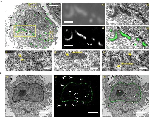
In this study, team members developed the first anti-xylate-fixed and Epon-embedded fluorescent protein, which remains fluorescent and has optical switch activity after TEM preparation, by optimizing single-molecule localization in ultrathin sections. Algorithm and imaging method, for the first time, the same layer super-resolution optical-electron microscopy correlation imaging of Epon post-fixed electron microscope samples was realized. The photoelectric correlation imaging method well preserves the subcellular structure such as mitochondria in the electron microscope image, and has the single molecule positioning accuracy of single-molecular localization super-resolution light microscopy imaging. The photo-association imaging of mitochondria and nuclear membrane was successfully achieved using this technical cooperation team (Figure). The fluorescent protein will be widely used in optoelectronic correlations such as nerve and brain science, which require continuous sectioning and 3D reconstruction of large-scale thick samples.
The instrument research and development team led by Xu Tao, an academician of the Chinese Academy of Sciences, has been working on the research and development of microscopic imaging equipment and technical methods in recent years. He has developed polarization single-molecule interference imaging, frozen single-molecular positioning imaging and super-resolution photoelectric fusion imaging systems. A new method of super-resolution microscopy imaging. Xu Pingyong's research group focuses on the development of a variety of probes for super-resolution imaging such as PALM/STORM, SOFI, SIM, etc., and develops new imaging methods based on the photochemical properties of probes such as Bayesian single-molecule super-resolution imaging method SIMBA Etc., to improve the spatiotemporal resolution of super-resolution imaging. This work is a collaboration between Xu Tao's research group and Xu Pingyong's research group. It is obtained in the field of super-resolution imaging after the PALM imaging probe mEos3.2 (Nature Methods, 2012) and the live cell super-resolution imaging method Quick-SIMBA (Nano Letter, 2019). Another important research result.
Xu Tao, associate researcher Zhang Mingqi and researcher Xu Pingyong are the co-authors of the article, Fu Zhifei, Peng Dingming, and Zhang Mingxi are the co-first authors.
The work was funded by the National Key Research and Development Program, the National Natural Science Foundation, the Chinese Academy of Sciences Pilot Project, the Chinese Academy of Sciences Research Equipment Development Project, and the Beijing Science and Technology Program.